
The patellar tendon is the fibrous band that connects the kneecap (patella) to the shinbone (tibia), just below the kneecap. It comes into play every time the leg straightens with force, when jumping, running, braking, or climbing stairs, which is why its injuries are common among people who play sports involving jumps and explosive movements.
This content is for informational purposes. Its goal is not to offer a treatment, but to explain what the patellar tendon is, why it is also called a ligament, which injuries affect it, and what the scientific evidence says about its capacity for repair.
In brief
- What it is: a band of fibrous tissue that runs from the lower end of the kneecap to a bony bump on the front of the shinbone called the tibial tuberosity.
- Tendon or ligament: these are two names for the same structure. It connects bone to bone, like a ligament, but it is the continuation of the quadriceps tendon, which is why it is called a tendon.
- What it does: it is part of the set of structures that straightens the knee, known as the extensor mechanism.
- Most common injuries: patellar tendinopathy, or “jumper’s knee,” and, much less often, a tear.
What the patellar tendon is and where it is located
The patellar tendon sits at the front of the knee, below the kneecap. It starts at the lower end of the kneecap, called the inferior pole, and attaches to the tibial tuberosity, the bony bump you can feel on the front of the shinbone, just below the knee. It is wide where it begins, matching the width of the kneecap, and narrows as it runs down to the tibia.
Behind it, a pad of fat known as Hoffa’s fat pad separates it from the membrane that lines the inside of the joint. The patellar tendon is not wrapped in a sheath but in a thin layer of loose tissue called the paratenon, just like the Achilles tendon.
With the leg straight and the muscle relaxed, the firm band you can feel between the kneecap and the shinbone is the patellar tendon.
Patellar tendon or patellar ligament?
Both names are used for the same structure, and each has a rationale. The difference lies in the criterion used to classify it.
A tendon connects a muscle to a bone, while a ligament connects two bones. The patellar tendon connects the kneecap to the tibia, that is, bone to bone, so it meets the definition of a patellar ligament. Its anatomical name in Latin is ligamentum patellae.
However, the kneecap is a bone located inside a tendon, the quadriceps tendon; in anatomy, this type of bone is called a sesamoid bone. From that point of view, the band that continues below the kneecap is the final portion of the quadriceps tendon before it reaches the tibia, which is why it is called a tendon. The word “patellar” comes from patella, the Latin name for the kneecap.
| Patellar tendon | Patellar ligament | |
|---|---|---|
| Criterion | Functional: it is the continuation of the quadriceps tendon | Anatomical: it connects two bones, the kneecap and the tibia |
| Origin of the name | Patella, the Latin word for kneecap | Ligamentum patellae, its Latin anatomical name |
| Is it the same structure? | Yes | Yes |
What the patellar tendon does
The patellar tendon is the final part of the set of structures that straightens the leg, known as the extensor mechanism of the knee. This system includes the quadriceps muscle, the quadriceps tendon, the patellar retinacula, the kneecap, the patellar tendon, and the tibial tuberosity.
Its job is to transmit the force of the quadriceps to the tibia so the knee can extend. That is why it works during everyday movements such as getting up from a chair or climbing stairs, as well as during jumps and sudden stops.
A healthy patellar tendon is very strong: the load needed to rupture it has been estimated at about 17.5 times body weight. When it does tear, it is usually because a sudden overload coincides with a tendon that was already weakened.
The patellar retinacula: neighboring structures that are not the tendon
The patellar retinacula are two fibrous reinforcements located on either side of the kneecap: one on the inner side (medial) and one on the outer side (lateral). They are thickenings of the fibrous layer that surrounds the knee joint, the joint capsule, and they blend with the edges of the patellar tendon.
Although they belong to the same extensor mechanism, they are not the tendon. For this reason, pain located on one side of the kneecap does not always come from the patellar tendon, and determining which structure is affected requires a clinical assessment and, when indicated, imaging tests.
Most common patellar tendon injuries
Patellar tendon injuries fall into two types that are worth telling apart: overuse injury, which develops gradually, and tears, which usually happen suddenly.
Patellar tendinopathy, or “jumper’s knee”
Patellar tendinopathy is an overuse injury that affects both professional and recreational athletes. It is not caused by a blow, and it is characterized by tendon pain that gradually increases. It is known as “jumper’s knee” because it affects athletes who perform jumps and explosive movements.
Reported risk factors include playing jumping sports, having a higher jump height, and training on hard surfaces. The pain is located in the tendon and increases when the knee is straightened against resistance.
The term “tendinopathy” is considered more accurate than “tendinitis” as a general description of tendon problems related to overuse.
Partial or complete tear
A patellar tendon tear, or rupture, is uncommon: it is estimated at about 0.68 cases per 100,000 people per year, less frequent than a quadriceps tendon rupture.
It usually happens during a sudden contraction of the quadriceps with the knee bent, for example when landing from a jump, and the area that most often tears is where the tendon meets the lower end of the kneecap. A previous injury, repeated overload, tendinopathy, and degenerative changes in the tendon increase the risk of rupture. An acute tear causes sudden pain and swelling below the kneecap after a specific event.
See also: Patellar tendon tear
| Patellar tendinopathy | Patellar tendon tear | |
|---|---|---|
| How it starts | Gradually, through repeated overload | Suddenly, after a specific event |
| How common it is | Common in jumping sports | Uncommon |
| Typical symptom | Pain that increases with load | Sudden pain and swelling below the kneecap |
| How they are related | It can weaken the tendon | It is more likely in a previously injured tendon |
Why the patellar tendon has difficulty healing
Tendinopathy is a difficult problem to treat: it usually requires prolonged management, and the response to treatment is often limited.
Part of the problem lies in how tendons heal. After an injury, the tissue repairs and remodels itself, but the resulting tissue does not fully recover the biochemical and mechanical properties of the original tendon. This helps explain why a previous injury is one of the factors that increase the risk of the patellar tendon eventually tearing.
See also: Truths and myths about mesenchymal cells in tendon regeneration
What the evidence says about the different approaches
The approach depends on the type of injury, how it has progressed, and each person’s characteristics.
Exercise therapy for tendinopathy. Stretching and strengthening exercises are part of the nonsurgical treatment of patellar tendinopathy. In a randomized clinical trial with 76 patients, a progressive loading program achieved better results than eccentric exercise at 24 weeks on a validated questionnaire of pain, function, and ability to play sports, although the difference in return-to-sport rates was not significant.
Surgery for a complete tear. For a complete tear, surgical repair is generally indicated, regardless of age or activity level. Nonsurgical management is reserved for partial tears in which the person can still straighten the knee.
Research with cultured mesenchymal cells (CMC). Cells become an advanced cell therapy medicinal product when they undergo substantial manipulation, for example when they are expanded in culture, under Regulation (EC) No 1394/2007. That is why CMC are manufactured in a clean room under GMP standards and require individual authorization from the AEMPS, the Spanish Agency of Medicines and Medical Devices.
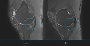
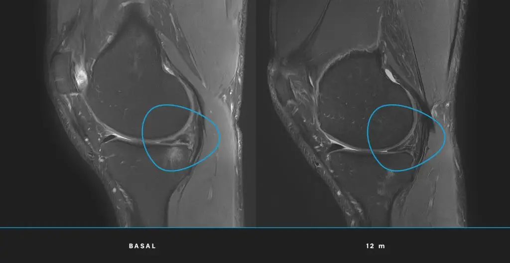
In an early-phase (phase 1/2), randomized, double-blind clinical trial authorized by the AEMPS, 20 athletes with chronic patellar tendinopathy, with a lesion larger than 3 mm in the part of the tendon closest to the kneecap and no response to nonsurgical treatments, received either CMC obtained from their own bone marrow or a biological control preparation. Both groups followed the same rehabilitation program. At 6 months, pain and function improved in both groups, with no significant differences between them. The difference was in the structure of the tendon: the CMC group showed significantly greater improvements on ultrasound and magnetic resonance imaging (MRI), and signs of normalization of the structure were seen only in that group (Rodas et al., Am J Sports Med, 2021, PMID 33783227).
At the 12-month follow-up, the control group also received CMC, so there was no longer a comparison group. Both groups showed clinical and structural improvement and no adverse effects were recorded, although in the group treated second, the improvement in pain during sports was not significant (Soler et al., Orthop J Sports Med, 2023, PMID 37711505). Because the sample was small, these results should be interpreted with caution and cannot be extrapolated to all tendon injuries.
See also: How to regenerate the patellar tendon with cultured mesenchymal cells
Not all patellar tendon injuries call for the same approach. Early tendinopathy, chronic tendinopathy with structural changes, and a complete tear are different situations, and the decision is made on an individual basis after a clinical assessment and imaging tests.
Frequently asked questions
Is the patellar tendon the same as the patellar ligament?
Yes. They are two names for the same structure. It is called a ligament because it connects two bones, the kneecap and the tibia, and a tendon because it is the continuation of the quadriceps tendon.
Where is the patellar tendon?
At the front of the knee, below the kneecap. It runs from the lower end of the kneecap to a bony bump on the front of the shinbone called the tibial tuberosity.
What is the patellar retinaculum?
The patellar retinacula are two fibrous reinforcements located on either side of the kneecap. They are part of the knee’s extensor mechanism along with the patellar tendon, but they are separate structures.
What is jumper’s knee?
It is the common name for patellar tendinopathy, an overuse injury of the patellar tendon that causes gradually increasing pain. It especially affects athletes who perform jumps and explosive movements.
What is the difference between patellar tendinopathy and a patellar tendon tear?
Tendinopathy develops gradually through repeated overload and causes pain that increases with load. A tear is uncommon, usually happens suddenly after a specific event, and causes sudden pain and swelling below the kneecap.
Does the patellar tendon heal on its own?
The tendon heals, but the tissue that forms does not fully recover the properties of the original tendon. In the case of tendinopathy, management is usually prolonged, and the approach depends on the type of injury and how it progresses.
Legal notice
Advanced cell therapy medicinal products with cultured mesenchymal cells (CMC) require a medical prescription and individual authorization from the AEMPS. This content is for informational purposes only and is not a substitute for medical advice. Clinical outcomes depend on multiple factors and cannot be extrapolated from one patient to another.